Generator
• High frequency, 2.5 kHz
• 7.5 kW full-wave
• Up to 120 kVp
• Up to 100 mA for radiographic film
exposures
• Boosted fluoro and Pulsed fluoro
capability
• Full power from standard outlet
120V/15A
• Patented energy buffer design
X-Ray Tube
• Rotating anode X-ray tube
• Focal spots: 0.3 mm and 1.0 mm
• Focal spot kW rating: 0.3 mm = 5.3 kW
and 1.0 mm = 41.0 kW
• Anode heat capacity: 300,000 H.U.
• Anode cooling rate: 70,000 H.U./min
• Housing heat capacity: 1,250,000 H.U.
• Anode diameter: 3 in
• Anode angle: 10˚
Collimator
• Two sets of opposing shutters
• Continuously variable and rotatable
• All functions remotely controlled from
the C-Arm control panel and optional
hand held remote
T.V. Camera
• 1 in Vidicon (high sensitivity)
• Progressive scan operation
• 360˚ motorized rotation
• On-screen orientation indicator
(real-time feedback without fluoro)
• Left-right image reversal
• Top-bottom image reversal
• Negative mode
• Bandwidth: 15 MHz
• Video signal: Standard RS-170 A 60 Hz,
525 line
• Aspect ratio: 4:3
• Computer controlled features:
– Dark current compensation
– Gain
– Blanking
– Camera iris
Video Monitors
• Dual 17 in Ultrabrite anti-glare
monitors
• 525 lines, 20 MHz
• Ambient room light sensor
• Remote brightness/contrast
controls
Image Processing
Expanded Surgical Platform (ESP)
• 60 image storage with last image hold
• 640 x 512 x 10 bit
• Frame averaging noise reduction
(low, medium, high)
• One-shot frame integration
(low, medium, high)
• MARS (motion artifact reduction
system)
• Variable edge enhancement (track ball)
• Digital window/level
• Real-time auto-histogramming
(auto window/level)
• Variable zoom and roam (track ball)
• Patient annotation keyboard |
Fluoroscopy Mode
• Focal spot: 0.3 mm
• kVp range: 40 – 120 kVp
• Maximum ripple: typically 1% at
120 kVp/5 mA
• mA range: 0.2 – 5.0 mA normal mode,
1.0 – 20 mA boost mode
• Auto and manual modes
• Continuous, one-shot or pulsed
operation
• ABS varies mA, kVp and camera gain
• User selectable ABS tables for
orthopedics, chest and low dose
applications
Pulsed Fluoroscopy Mode
• Focal spot: 0.3 mm
• kVp range: 40 – 120 kVp
• mA range: Up to 60 mA
• Pulse rate: 1, 2, 4, or 8 pulses per
second
• Pulse width: 30 or 50 milliseconds
• Camera operates in progressive scan
mode
• Computer controlled camera iris, mA,
kVp and camera gain
• Anatomical markers
• On-line help menus
• Chole mode (up to 20 mA)
• 4 F/S digital disc (optional)
Vascular Module
Includes all Expanded Surgical Platform
(ESP) features plus adds:
• Real-time subtraction
• Roadmapping
• Peak opacification
• Re-registration (track ball)
• Variable landmarking (track ball)
• Mask save/recall
4 F/S Digital Disc (optional)
• Record rate: 1, 2 or 4 Frames/Sec
• Record time: 20, 10 or 5 minutes
• Play mode: 1, 2 or 4 Frames/Sec
• Control: Instant image access
(track ball)
• Synchronized to pulse with generator,
T.V. camera and image processor
Hardcopy Options
• Radiographic film capability
• Thermal printer
System Control
• Entire system is computer controlled
• Software upgradable
• Main control panel pivots right-to-left
for user convenience
• Multi-function infrared remote control
(optional)
• Hand held X-ray remote control
(optional)
• Multi-function footswitch |
Radiographic Mode
• Focal spot: 0.3 mm or 1.0 mm
• Focal spot automatically selected
• mAs range: 1 – 300 mAs
• kVp range: 50 – 120 kVp
• Cassette holder (optional) 10 in x 12 in
(24 cm x 30 cm) 9 in I.I.
Video Imaging System
Image Intensifier
• Tri-mode 9 in/6 in/4.5 in image
intensifier
• Central resolution (typical):
4.5 in: 54 lp/cm
6 in: 48 lp/cm
9 in: 41 lp/cm
• Peripheral resolution at 70% radius
(typical):
4.5 in: 50 lp/cm
6 in: 46 lp/cm
9 in: 40 lp/cm
• Contrast Ratio: 30:1 (typical)
• DQE: 65% (typical at 1.7 Hz)
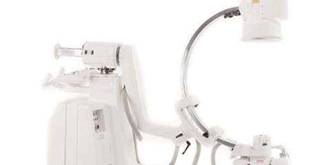
Mechanical
• Source to image distance: 36.18 in
(919 mm)
• Free space in arc: 27.2 in (691 mm)
• Depth of arc: 23.3 in (592 mm)
• Arc orbital movement: 111˚
• Left/right wig-wag scan: ±11˚
• Vertical travel: 18 in motorized
(457 mm)
• Horizontal travel: 8 in (203 mm)
• L-Arm rotation: ±185˚ motorized
• Reversible C-Arm: 180˚ manual flip-flop
C-Arm Dimensions In compacted position:
• Length: 74.5 in (1892 mm)
• Width: 34 in (864 mm)
• Height: 65.8 in (1671 mm)
Workstation Dimensions
• Depth: 27 in (686 mm)
• Width: 32 in (813 mm)
• Height: 65 in (1651 mm)
Electrical
• Input power (50 Hz or 60 Hz):
115 V/15A
230 V/12A
100 V/20A |